The CPC provides pathology support for research projects involving animal models. This includes gross and microscopic pathology, clinical pathology, special imaging, photography and data analysis. The CPC provides service to investigators both within and outside Iowa State University and can assist with a broad range of preclinical studies in a wide range of species. Studies that CPC has completed include toxicology, vaccine studies, infectious disease, nutritional physiology, neoplastic disease, musculoskeletal disease and developmental disorders.
Contact information
Please feel free to contact the facility if you have any question regarding this service or how it could benefit your current or future projects.
Rachel Phillips
Assistant Scientist II
rlp79@iastate.edu
515-294-0953
or fill out this request for service form and email to rlp79@iastate.edu
Pathology
- Gross examination
- Tissue collection
- Gross lesion scoring
- Histopathology
- Microscopic lesion scoring
- Special stains
Clinical Pathology
- CBC and serum biochemical analysis
- Cytologic examination
- Urinalysis
Special Imaging
- Immunohistochemistry
- Fluorescence
- Chromogenic
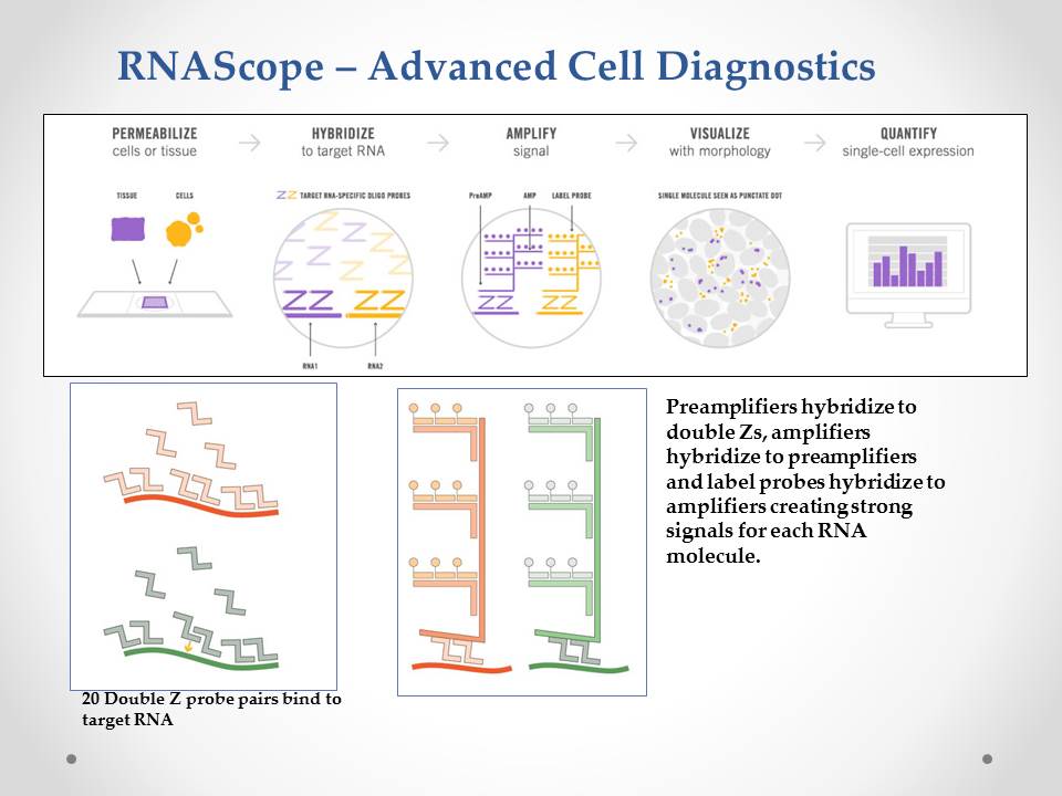
- In Situ Hybridization – RNAscope
- Hyperspectral microscopy
Photography
- Gross and microscopic images
- Fluorescent and chromogenic IHC
- Formatting for publication
Data Analysis and Report Generation
- HALO Image Analysis software
- Morphometrics
Important Note: This is not a GLP laboratory. If any of the data derived from testing will be submitted to FDA, CVB, EPA, NCI or another federal agency, you are required to notify us of the agency and present the applicable regulations to our laboratory. Data from CPC may not be submitted for safety/efficacy to FDA, CVB, EPA, NCI, or other federal agencies.
Featured Service
In Situ Hybridization - RNAscope


This is a new method for in situ hybridization that yields a strong, specific, readable signal (see figures below) using formalin fixed / paraffin embedded or frozen tissues. Species specific probes can be designed for multiple species including bovine, porcine, canine, equine and murine. Please contact us to find out if this technology may be helpful in your research project.

Mission: Provide high quality pathology support to researchers using animal models.
Contact Information:
Rachel Phillips
Assistant Scientist II
rlp79@iastate.edu
515-294-0953
Amanda Fales-Williams
Faculty Contact
fales@iastate.edu
515-294-7445
