An inordinately large number of cases of epizootic hemorrhagic disease (EHD) have been reported in the Midwestern United States this year. Odd in of itself due to drought conditions. Epizootic hemorrhagic disease viruses (EHDV) are widespread in white-tailed deer and periodically cause serious epidemics in wild populations as well as affecting farmed deer populations. EHD can also cause disease in cattle. Lesions mimic those observed with BVDV, plant photosensitization, adenovirus, Blue Tongue virus and Foot and Mouth Disease. As such, animals with lesions should be reported to the federal area veterinarian in charge as well as the state veterinarians’ office.
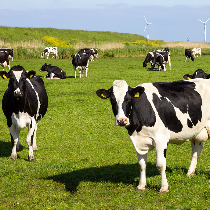
In the U.S., EHD in cattle is typically uncommon, rarely fatal, and usually associated with an epidemic in deer as has been seen this year.
The epizootic hemorrhagic disease viruses belong to the genus Orbivirus, family Reoviridae. Ten serotypes of EHDV are known worldwide. The viruses of the EHDV serogroup are transmitted by biological vectors, usually biting midges in the genus Culicoides. In North America, C. variipennis is the major vector. Some species of gnats and mosquitoes can also transmit EHDV. Infected deer can be viremic for up to 2 months. The incubation period for epizootic hemorrhagic disease in deer is 5 to 10 days.
Deer
Three syndromes may be seen in deer. Peracute disease is characterized by high fever, anorexia, weakness, respiratory distress, and severe and rapid edema of the head and neck. Swelling of the tongue and conjunctivae is common. Deer with the peracute form usually die rapidly, typically within 8-36 hours. Some animals may be found dead with few clinical signs. In the acute form these symptoms can also include extensive hemorrhages in many tissues including the skin, heart, and gastrointestinal tract. There is often excessive salivation and nasal discharge, which may both be blood-tinged. Animals with the acute form can also develop ulcers or erosions of the tongue, dental pad, palate, rumen, and omasum. High mortality rates are common in both the peracute and acute forms. The chronic form of the disease results in deer being ill for several weeks but gradually recover. After recovery, these deer sometimes develop breaks or rings in the hooves caused by growth interruption and may become lame. In severe cases, animals slough the hoof wall or toe; some of these deer may be found crawling on their knees or chest. Deer with the chronic form may also develop ulcers, scars, or erosions in the rumen; extensive damage to the lining of the rumen can cause emaciation even when there is no shortage of food.
Cattle
Disease in cattle is characterized by fever, anorexia, and difficulty swallowing. The swallowing disorders are caused by damage to the striated muscles of the pharynx, larynx, esophagus and tongue, and may lead to dehydration, emaciation, and aspiration pneumonia. Edema, hemorrhages, erosions, and ulcerations may be seen in the mouth, on the lips, and around the coronets. The animals may be stiff and lame, and the skin may be thickened and edematous. Abortions and stillbirths have also been reported in some epidemics. Most EHDV infections in cattle appear to be subclinical. Typical symptoms include fever, oral ulcers, salivation, lameness associated with coronitis, and weight loss. In pregnant cows, the fetus may be resorbed or develop hydranencephaly if it is infected between 70 and 120 days of gestation. Deaths are uncommon with the North American strains of EHDV; however, some animals may be lame and unthrifty for a prolonged period. Epizootic hemorrhagic disease has not been reproduced in experimentally infected cattle; although animals become viremic, they remain asymptomatic.
Other species
Sheep can be infected by EHDV but rarely develop symptoms. Experimentally infected goats do not become viremic.
Most outbreaks of EHD occur in late summer or early autumn. The onset of freezing weather usually stops the appearance of new cases, but hoof sloughing can be seen throughout the year.
Among the Cervidae, EHD is most severe in white-tailed deer. In this species, the morbidity and mortality rates may be as high as 90%. Severity of the disease varies from year to year. It also varies with the geographic location. In the southeastern U.S., most cases are mild and the mortality rates are low. In the Midwest and Northeast, EHD typically recurs each year, but can vary from a few scattered cases to severe epizootics with high mortality rates. This variability is thought to be caused by many factors including the abundance and distribution of the insect vectors, the EHDV serotype, existing herd immunity, and genetic variations in the susceptibility of the host. Surviving deer develop long-lived neutralizing antibodies. Nearly 100% of the deer population can be seropositive in some regions.
Samples to collect
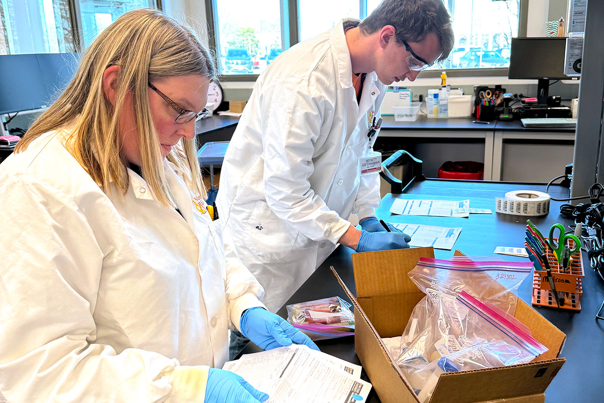
Preferred tissues in deer are spleen, lymph node, and unclotted whole blood in EDTA or heparin. Other useful samples may include serum (for serology), liver, and lung. Both fresh and fixed tissues should be collected if possible. Samples for virus isolation should be transported under refrigeration. For suspect cases in cattle, blood should be collected into anticoagulant (calcium citrate, EDTA, or heparin) and sent chilled for virus isolation or RT-PCR. Paired serum samples should also be collected if possible. Additionally because of other differentials involved fluid from intact vesicles as well as aggressive swabs or biopsy samples near lesions are also suggested.
Should questions arise please feel free to contact any of the diagnosticians at the ISU-VDL (515-294-1950).
More information on EHD may also be found on the Center for Food Security & Public Health web site.
http://www.cfsph.iastate.edu/DiseaseInfo/disease.php?name=epizootic-hemorrhagic-disease&lang=en